From Surface to Sample: Investigating how everyday surfaces contribute to microbial contamination in a controlled laboratory environment
From Surface to Sample
Investigating how everyday surfaces contribute to microbial contamination in a controlled laboratory environment
Authors: Avedikian, N; Johnston, W.; Rodriguez, J.; Leano, S.
Abstract:
Microbial contamination stands as an important concern in biotechnology labs as unwanted bacteria and fungi have the ability to alter experimental accuracy and hinder controlled research environments. Although Biosafety Level 2 (BSL-2) labs are designed to reduce contamination through constant sanitation procedures, controlled airflow, and specialized sterilizing equipment, microorganisms can still be introduced into these environments through frequently touched surfaces and objects that are exposed to outside environments. Understanding which surfaces are most likely to introduce these harmful contaminants is paramount for improving lab safety and contamination-control practices. While preventing contamination is accounted for by many labs, there has been ample research focused on comparing contamination levels among common surfaces within a single BSL-2 lab. Surfaces such as door handles, chairs, footwear, and lab equipment serve as pathways through which microorganisms and bacteria enter controlled environments; thus, bringing in unwanted contaminants and negatively affecting bio-safety. By identifying these potential sources of contamination, scientists can effectively target their sanitation efforts and environmental monitoring programs in controlled lab environments.Furthermore, the purpose of this study was to investigate whether frequently touched and exposed surfaces could act as sources of bacterial and fungal contamination within a BSL-2 lab. Samples were collected from a fire extinguisher, dry water bath, biosafety cabinet, exterior door handle, lab chair, and the bottom of a lab worker’s shoe using sterile swabs. A closed control plate exposed to the lab environment for twenty minutes to serve as comparison to the other samples. However, while some samples were collected from commonly contacted surfaces within the lab, samples such as the fire extinguisher, shoe sole, and exterior door handle served as proxies for samples of contamination such as dust, contaminants around the lab’s floors, and bacterial buildup from constant contact. Next, samples were cultured on nutrient agar plates and incubated at 37°C. Microbial growth was evaluated after 24 and 96 hour increments by observing colony formation, estimating number of colonies formed, and comparing the various present bacteria and fungi growing on the sampled surfaces. It was hypothesized that surfaces exposed to greater human contact would exhibit higher levels of microbial growth than routinely sterilized lab equipment.
Ultimately, results indicated that microbial growth was present on multiple surfaces inside and outside the lab, including both bacterial and fungal colonies. The fire extinguisher, shoe sole, and lab chair exhibited the highest levels of contamination, while the dry water bath showed comparatively lower contamination. Growth was also observed on the control plate, suggesting the possible presence of airborne microorganisms within the lab environment. These results support the hypothesis that surfaces with higher contact and exposure to sources of bacteria may serve as key pathways for introducing harmful microorganisms into controlled lab settings. All in all, this study highlights the value of environmental monitoring while demonstrating the importance of maintaining effective sanitation and contamination-control practices within controlled lab spaces.
Introduction:
Bacteria are found on nearly every surface in the environment and can be studied through lab cultures to better understand patterns of contamination and microbial growth (Maier & Pepper, 2015). In controlled environments like laboratories, cross-contamination can occur when bacteria are transferred from frequently contacted surfaces into sterile areas. Since contamination can affect both lab-safety and experimental precision, maintaining clean lab environments is a very important aspect of scientific procedures. Although biological safety labs are designed to reduce contamination through controlled airflow, filtration systems, and constant cleaning procedures, studies have shown that particle movement and bacterial growth can still influence contaminant distribution within lab environments (Hu, 2015).
While controlling contamination in lab settings is regarded to an extent, relatively few investigations compare bacterial contamination among commonly encountered surfaces within a single Biosafety Level 2 (BSL-2) laboratory. Frequently touched surfaces, lab equipment, and objects exposed to outside environments can serve as pathways through which microorganisms enter controlled lab spaces. Understanding which surfaces carry the greatest amount of contamination can provide insight into potential sources of bacterial transfer and help polish environmental monitoring practices.
The purpose of this study is to evaluate bacterial growth on multiple environmental and lab surfaces, including a fire extinguisher, a dry water bath, a biosafety cabinet, an exterior door handle, a lab chair, and the bottom of a BSL2 worker’s shoe. The control plate, exposed to the lab environment for twenty minutes, was also included for comparison. Samples were collected using sterile swabs, cultured on LB agar plates, and analyzed for bacterial colony growth in each sample. It was hypothesized that surfaces exposed to frequent human contact or the outside environment, particularly the shoe and door handle, would exhibit greater bacterial growth than surfaces located within controlled lab work areas. The results from this investigation give insight into where potential contamination pathways lie. Whether it be inside or outside the lab is a question that arises from this investigation as it highlights the significant importance of maintaining effective contamination control practices in a controlled lab setting.
Methods:
Sample Collection:
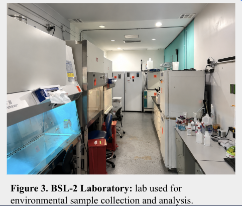
Samples were collected from seven locations inside and outside the lab that were associated with the lab’s environment. These locations included a fire extinguisher, dry water bath, biosafety cabinet, exterior door handle, laboratory chair, the bottom of a laboratory worker’s shoe, and a control sample. Sterile swabs were used to collect microbial samples from each surface. The closed control plate was exposed to the laboratory environment for twenty minutes without direct swabbing to evaluate possible airborne contamination.
Culturing/Incubation:
After all samples were collected with the sterile swab, each sample was streaked onto a labeled LB agar plate using sterile technique. The inoculated plates were labeled according to their sampling location and placed in an incubator at 37°C. Plates remained in the incubator for increments of 24 and 96 hours where a new set of samples were taken for the 96 hour investigation in order to allow microbial growth and colony development in each sample.
Data Collection/Analysis:
Microbial growth was observed and recorded after 24 hours and again after 96 hours of incubation. Observations included colony formation, colony appearance, and the presence of bacterial or fungal growth. Samples were compared to determine differences in contamination levels among the tested surfaces. Microscopic observations were conducted at both time points to further evaluate microbial diversity more closely as colony characteristics and contamination patterns were more evident under the magnified lens.
Results:
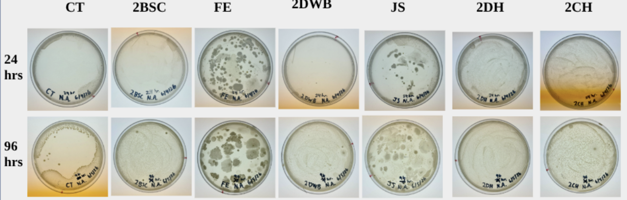
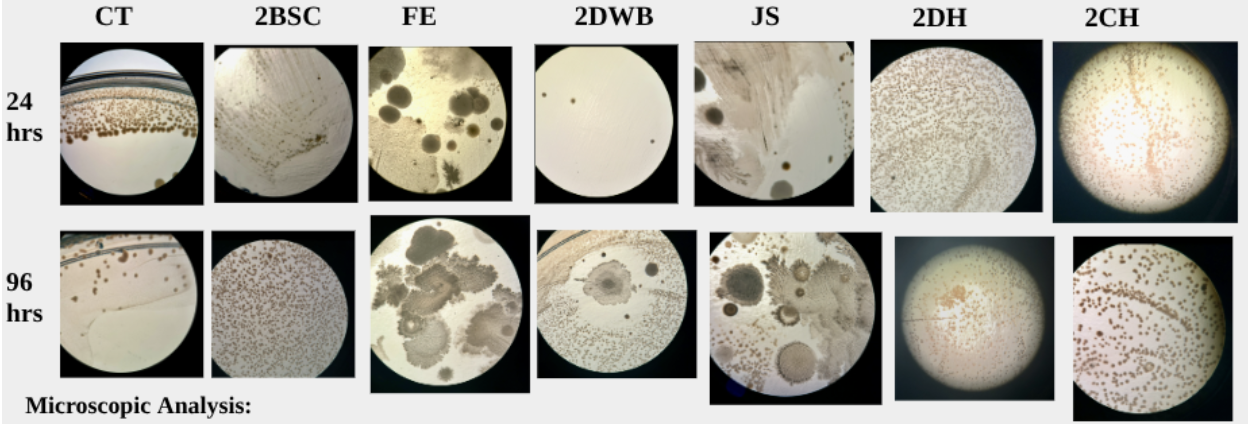
Microbial growth varied significantly among sampled surfaces, with some locations in the environment exhibiting substantial colony formation than others after the full 24 and 96 hours of incubation. Standard observations of the agar plates revealed differences in microbial growth after 24 and 96 hours of incubation. The fire extinguisher and shoe samples exhibited the most visually diverse growth, with large irregular colonies increasing over time. In contrast, the dry water bath and biosafety cabinet displayed lower contamination levels. Growth on the control plate indicated the presence of airborne microorganisms. While visual observations provided an initial assessment, microscopic analysis offered a more accurate evaluation of microbial diversity and bacterial and fungal growth, making it the primary method for assessing contamination among the sampled surfaces. Furthermore, the majority of the seven sampled surfaces exhibited abundant growth of small, circular bacterial colonies that resembled E. coli morphology and produced a characteristic odor commonly associated with E. coli cultures. Similar colonies were observed on the control plate, dry water bath, door handle, laboratory chair, and biosafety cabinet samples. These colonies increased in abundance between the 24 and 96 hour observations, suggesting that active microbial growth occurred under the incubation conditions. The presence of these bacterial colonies indicates that microorganisms can persist on both laboratory equipment and frequently contacted surfaces, even within a controlled BSL-2 environment that is sterilized regularly (CT, 24 & 96; 2BSC, 24 & 96; 2DWB, 24 & 96; 2DH, 24 & 96; 2CH, 24 & 96).
However, several samples exhibited microbial growth patterns that differed significantly from the other surfaces. The fire extinguisher sample displayed multiple unusually large, irregular colonies with varying sizes, shapes, and microbial characteristics, indicating a highly diverse microbial population. In addition to bacterial colonies, fungal growth was observed, including dark colonies consistent with black mold morphology. The shoe sample also exhibited significant microbial diversity, including both bacterial and fungal colony morphologies. Since footwear routinely comes into contact with floors and external environments, the shoe sole likely served as a carrier for transporting microorganisms into the laboratory. These observations support the hypothesis that environmentally exposed surfaces may serve as major pathways for contamination within controlled laboratory environments (FE, 24 & 96; JS, 24 & 96).
Among all samples, the fire extinguisher and shoe sole presented the greatest diversity of microbial growth, while the door handle and laboratory chair exhibited the highest abundance of small bacterial colonies. These findings suggest that both environmental exposure and frequent human contact contribute to contaminants that enter the BSL-2 laboratory, highlighting the importance of targeted sanitation and environmental monitoring practices (FE, 96; JS, 96; 2DH, 96; 2CH, 96).
Discussion:
Analysis:
The results of this investigation indicated that microbial contamination was present on multiple surfaces inside and outside the BSL-2 lab environment. Both bacterial and fungal growth were observed on several agar plates, indicating that microorganisms can persist on commonly encountered surfaces despite routine sanitation procedures. The fire extinguisher, laboratory chair, and bottom of the laboratory worker’s shoe exhibited the greatest levels of contamination, while the dry water bath displayed comparatively lower microbial growth. These findings suggest that surfaces exposed to frequent human contact or environmental exposure may serve as major pathways for introducing microorganisms into controlled laboratory settings.
The results generally supported the original hypothesis that surfaces with greater contact and exposure would exhibit higher levels of microbial growth than routinely maintained lab equipment. The significant growth observed on the shoe sole was expected because footwear regularly comes into contact with floors and outside environments, allowing microorganisms to be transported into the lab. Similarly, the high contamination levels observed on the fire extinguisher and laboratory chair suggest that rarely sanitized or frequently touched surfaces may accumulate microorganisms over time. Growth on the control plate also suggests that airborne microorganisms may contribute to contamination within the lab environment.
These findings highlight the importance of environmental monitoring in biotechnology labs. While BSL-2 labs are designed to minimize contamination through controlled procedures and sterilization practices, the results demonstrate that potential contamination sources still exist. Identifying surfaces with higher levels of microbial growth can help labs prioritize cleaning efforts and improve contamination-control strategies, ultimately improving lab safety and experimental accuracy.
Limitations/Modifications:
Several limitations should be considered when interpreting the results. Only a single sample was collected from each surface, which may not fully represent contamination levels throughout the laboratory. In addition, microbial identification was limited to visual observations of colony morphology rather than species-level analysis. Future studies could increase the number of samples collected, investigate additional surfaces, and use molecular or biochemical techniques to identify specific microorganisms. These modifications would provide a more complete understanding of contamination pathways and help develop more effective contamination-control strategies.
Conclusion:
This investigation uncovered the measurable bacterial contamination on several commonly exposed surfaces within a BSL-2 laboratory. The presence of bacterial colonies on frequently contacted surfaces supports the fact that microorganisms can still become introduced into controlled lab environments despite measures like air filtration set to control contamination. These findings highlight the importance of constant sanitation throughout sterile lab environments: including maintaining appropriate hygiene in the lab, monitoring of surfaces, and constant attention to detail. Furthermore, understanding where contamination is present and likely to occur can help improve lab safety and reduce risk of unwanted contamination and microbial transfer during scientific analysis.
References:
Hu, S.-C. (2015, August 11). Validation of cross-contamination control in biological safety cabinet for biotech/pharmaceutical manufacturing process. Springer Nature Link. Retrieved June 4, 2026, from https://link.springer.com/article/10.1007/s11356-015-5091-5
Maier, R. M., & Pepper, I. L. (2015). Chapter 3 - Bacterial Growth. ScienceDirect. Retrieved June 4, 2026, from https://www.sciencedirect.com/science/chapter/edited-volume/abs/pii/B978012394626300003X